

Cruciate Ligament in Dogs: Cranial Cruciate Ligament Disease (CCL/ACL) Complete UK Owner's Guide
If your dog has suddenly started limping on a back leg, or your vet has mentioned a possible cruciate ligament problem, you are likely feeling worried and searching for answers. This guide explains everything UK dog owners need to know about cranial cruciate ligament disease - from first signs through to diagnosis, treatment, surgery, recovery and long-term care. It is written to help you understand what is happening, what to expect, and how to make the best decisions alongside your veterinary team.
Quick answer: cruciate ligament rupture in dogs
The cranial cruciate ligament is a tough band of tissue inside your dog's knee that prevents the shin bone from sliding forward relative to the thigh bone. It is one of two cruciate ligaments in each stifle joint, and it is the one that almost always fails. In human medicine, the equivalent structure is called the anterior cruciate ligament, which is why many vets still use the term "ACL injury" when explaining the condition to owners. You may also hear this described as a torn acl in dogs.
Cranial cruciate ligament disease is the single most common knee injury causing back leg lameness in UK dogs. It affects all breeds and sizes, though certain breeds and overweight dogs are at higher risk.
In most dogs, this is not a sudden sports injury. The ligament fibres weaken gradually through a process of chronic degeneration over months or years. Eventually, the damaged ligament fails - often during ordinary activity like chasing a ball or jumping off the sofa - leaving the knee unstable and painful.
Treatment usually involves cruciate ligament surgery combined with structured rehabilitation, although some smaller or medically fragile dogs may be managed conservatively under veterinary guidance. The reassuring news is that with timely veterinary care, appropriate weight management and good rehabilitation, most dogs regain comfortable, active lives.
When to seek urgent veterinary care for a suspected cruciate injury
Some signs of cruciate ligament injury need same-day or emergency veterinary assessment. Cruciate rupture can look very similar to fractures, dislocations or hip problems, and only a professional examination can distinguish between them.
Seek urgent veterinary care if your dog shows any of the following:
- Sudden non-weight-bearing lameness - carrying a back leg completely off the ground
- Yelping, crying or refusing to move after a jump, turn or fall
- Visible swelling or heat around the knee joint
- Obvious deformity of the leg
- Inability to stand on one or both back legs
- After a road traffic accident, a fall from height, or a dog fight, limping may be caused by multiple injuries including fractures, internal bleeding or soft tissue damage. Never assume "it's just the cruciate" without professional assessment.
If your dog remains severely lame after 24 hours of rest, or the lameness worsens rather than improves, contact your vet without delay. For out-of-hours emergencies, call your practice's emergency number, describe the signs clearly, and follow their advice on whether to attend immediately. Pain relief and stabilisation are usually available quickly once the dog is assessed.
What and where are the cruciate ligaments in dogs?
Each of your dog's back legs contains a knee joint - veterinary surgeons call it the stifle joint. Inside each stifle sit two cruciate ligaments: the cranial cruciate and the caudal cruciate. They are called "cruciate" because they cross over each other inside the joint, forming an X shape.
These ligaments connect the femur (thigh bone) to the tibia (shin bone) and are comparable to the ACL and PCL in human knees. Together, they provide knee stability by preventing the tibia from sliding forwards or backwards, limiting excessive internal rotation, and stopping the joint from hyperextending when the dog walks, runs or jumps.
The cranial cruciate ligament (CCL) is the structure almost always affected in canine cruciate ligament disease. Caudal cruciate injuries are rare. When vets refer to "cruciate disease" or "cranial cruciate rupture," they are describing a common pattern of progressive degeneration and eventual rupture of this specific ligament.
The stifle also contains two C-shaped cartilage pads called menisci, which act as shock absorbers between the femur and tibia. These menisci, along with the smooth articular cartilage covering the bone surfaces, depend on an intact cruciate ligament to function normally. When the cruciate fails, these structures become vulnerable to damage.
How the cranial cruciate ligament works – simple knee anatomy
Understanding how the dog's knee is built helps explain why cruciate injuries cause such significant problems. Think of the stifle as a hinge joint held together by several key structures.
The main components include:
- Femur - the large thigh bone above the joint
- Tibia - the shin bone below the joint
- Patella - the kneecap, which glides in a groove on the front of the femur
- Joint capsule - a fibrous sleeve enclosing the joint, filled with lubricating synovial fluid
- Cruciate ligaments - the cranial and caudal crosses inside the joint
- Collateral ligaments - bands on the inner and outer sides preventing sideways movement
- Menisci - two cartilage cushions sitting between femur and tibia
In dogs, the top of the tibia - the tibial plateau - is naturally sloped. When the dog bears weight, this slope causes the femur to push backwards, which in turn drives the tibia forwards (a force called "tibial thrust"). A healthy ligament constantly resists this thrust, keeping the joint stable.
When the cranial cruciate ligament is intact, movement is smooth: the joint flexes and extends without the shin bone shifting forward, and the dog bears weight evenly and comfortably. When the ligament is stretched, partially ruptured or completely torn, the tibia can slide forward relative to the femur - creating the instability, pain and damage that characterise cruciate disease.
Why cruciate ligament disease happens in dogs
One of the most important things to understand is that most canine cruciate ligament rupture is not caused by a single dramatic accident. Unlike the classic human acl injury - a footballer twisting their knee during a match - the canine version is overwhelmingly a disease of gradual degeneration.
Microscopic damage accumulates in the ligament fibres over time. Collagen becomes disorganised, inflammation develops within the joint, and the ligament grows weaker, thicker and less elastic. Eventually, everyday activities - turning sharply in the garden, hopping out of the car, or even just trotting across a field - cause the weakened structure to fail. This surprises many owners who expect a visible traumatic event.
Several factors contribute to this degenerative process:
- Genetics - inherited differences in collagen structure and joint conformation play a major role. Research in Labrador Retrievers identified approximately 99 genetic loci associated with cruciate rupture, with heritability estimates around 0.48–0.49.
- Body weight and obesity - excess weight increases mechanical load on the ligament with every step
- Conformation - a steep tibial plateau angle, straight stifles, or abnormal limb alignment amplify shear forces
- Hormonal and immune influences - inflammatory processes within the joint may accelerate degeneration
- Age - the ligament weakens with time; most ruptures occur in middle-aged to older dogs
Once this degenerative process is established, both knees are typically affected to some degree. This explains why so many dogs eventually injure the opposite knee - it is not bad luck, but the same underlying cruciate disease progressing in both joints.
Which dogs are most at risk of cruciate ligament rupture?
Cranial cruciate ligament disease can affect any breed or crossbreed, but it is significantly more common in certain dogs. A VetCompass study of over 170,000 dogs in England found an overall prevalence of 0.56%, with clear breed, size and lifestyle patterns.
Higher-risk breeds commonly seen in UK practice include:
|
Risk category |
Example breeds |
|---|---|
|
Higher risk |
Labrador Retriever, Golden Retriever, Rottweiler, Boxer, Newfoundland, West Highland White Terrier, Bichon Frise |
|
Commonly affected |
Staffordshire Bull Terrier, Yorkshire Terrier, German Shepherd, Akita, Mastiff breeds |
|
Can still be affected |
Any breed or crossbreed, including small and toy breeds |
Medium-to-large, young-to-middle-aged dogs (approximately 2–8 years old) are most commonly affected, with the average age at diagnosis around 7.4 years. However, cruciate disease also occurs in older and smaller dogs.
Modifiable risk factors make a significant difference. Overweight dogs face substantially higher risk, and rapid weight gain in young large breed dogs is particularly concerning. "Weekend warrior" exercise patterns - sedentary during the week followed by intense weekend activity - may also contribute.
The evidence around neutering is important but complex. Some studies suggest that dogs of certain breeds neutered before 12 months of age have a higher risk of cruciate rupture, possibly due to altered hormonal influences on ligament and joint development. However, the relationship between neutering, obesity, and hormonal effects remains under active investigation. Neutering decisions should always be individualised through discussion with your veterinary surgeon.
Common signs and symptoms of cruciate ligament injury
Cruciate disease can look quite different from dog to dog, and the early signs are often surprisingly subtle. Understanding what to watch for can help you seek veterinary advice before the problem worsens.
Typical acute signs of a complete rupture:
- Sudden hindlimb lameness during or after exercise
- "Toe-touching" on the ground - barely putting weight on the affected leg
- Holding one back leg up entirely
- Reluctance to jump into the car or onto furniture
- Yelping or crying when the knee is moved or twisted
Chronic or early signs of developing cruciate disease:
- Intermittent lameness on the affected limb that improves with rest but returns after activity
- Stiffness after lying down, especially on cold mornings
- Difficulty on stairs or reluctance to climb
- "Bunny-hopping" with both back legs when running
- Sitting with one leg stuck out to the side (sometimes called a "sloppy sit")
Physical changes owners may notice include reduced muscle mass on the thigh of the injured leg, thickening or puffiness around the knee, or a soft clicking sensation when the leg is flexed.
It is worth remembering that dogs rarely vocalise knee pain in the way we might expect. Ongoing lameness or reluctance to use a leg almost always indicates discomfort, even if the dog seems otherwise cheerful and is still wagging its tail.

How vets diagnose cranial cruciate ligament disease
Diagnosis of cranial cruciate ligament disease is based on a combination of clinical history, physical examination and imaging.
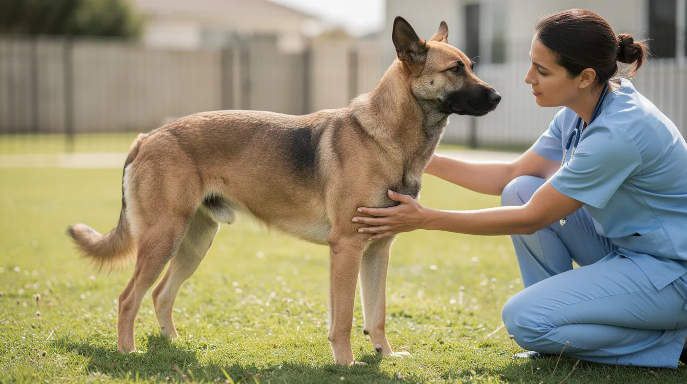
Your vet will typically start by watching your dog walk and trot, looking for lameness patterns, weight shifting, and how the dog stands at rest. They will then feel the stifle for swelling, warmth and pain, and gently test the range of motion.
Two specific manual tests help detect cruciate instability:
- Cranial drawer test - the vet holds the femur still and tries to slide the tibia forward. In a normal joint with an intact cruciate ligament, there is no movement. A positive result (forward sliding) strongly suggests cruciate rupture.
- Tibial compression test - the vet flexes the hock (ankle) while stabilising the femur, which loads the stifle and reveals tibial thrust if the cruciate is torn.
Many dogs tense their muscles during examination, which can mask instability. For this reason, sedation or light anaesthesia is often necessary for a reliable assessment. This also allows a less stressful experience for the dog.
X-rays (radiographs) are used to rule out fractures, measure the tibial plateau angle, assess joint swelling, and look for signs of osteoarthritis. The ligament itself does not appear on X-ray, so diagnosis relies on the clinical tests combined with imaging findings.
In complex cases - particularly where partial tears, a damaged meniscus, or meniscal injury is suspected - advanced imaging such as MRI or CT may be recommended, and arthroscopy can allow direct visualisation inside the joint.
What happens inside the knee when the cruciate ligament ruptures?
A ruptured cranial cruciate ligament creates both a mechanical and an inflammatory problem inside the joint.
The immediate effect is loss of stabilisation. Without the cranial cruciate to hold it in place, the tibia slides forward with every step, and abnormal rotation occurs within the joint. This causes micro-trauma to the cartilage surfaces and menisci with each weight-bearing movement, even during gentle dog walks.
Within days, synovial inflammation develops. The joint lining becomes irritated and produces excess fluid, causing visible swelling and increasing knee pain. This inflammatory process further degrades cartilage and remaining ligament tissue, creating a cycle of damage.
The medial meniscus is particularly vulnerable. When the unstable femur grinds across the tibial surface, the meniscus can become pinched or torn - a meniscal tear that often causes a sudden, dramatic worsening of lameness and sometimes an audible click. A damaged meniscus may need to be addressed during surgery.
Over weeks to months, the body responds with new bone formation around the joint margins, thickening of the joint capsule, and progressive arthritis development. Even dogs that appear to "cope" with a ruptured ligament are developing irreversible changes within the joint.
Stabilising the joint - whether surgically or, in selected cases, conservatively - aims to reduce this excessive movement and slow further arthritis development. However, no treatment can fully reverse damage that has already occurred.
Non-surgical (conservative) management of cruciate ligament injuries
Conservative or medical management focuses on pain relief, controlled exercise and muscle strengthening rather than physically altering the joint. It is usually considered when surgery is not feasible or in specific clinical situations.
Dogs that may be candidates for non-surgical care include:
- Small dogs (generally under 10–15 kg) with partial tears or mild instability
- Older dogs with serious concurrent medical conditions that increase anaesthetic risk
- Dogs whose owners decline surgery after a fully informed discussion with their vet
The core components of conservative management involve:
- Strict rest initially (typically 4–6 weeks of severely restricted activity)
- Gradual, controlled lead exercise, increasing incrementally under veterinary guidance
- Weight loss where the dog is overweight
- Anti inflammatory medications and pain management tailored to the individual
- Physical therapy and, where available, hydrotherapy to rebuild muscle mass
- Regular veterinary reviews to adjust the treatment plan
Owners should have realistic expectations. Recovery through medical management is typically slower than after surgery, some degree of residual lameness is common, and arthritis will continue to progress in the unstable joint. In VetCompass UK data, approximately 24% of cruciate cases were managed non-surgically, suggesting this is a real and valid pathway for selected dogs.
It is important to dispel the myth that "two weeks of rest and back to normal" is realistic for true cruciate ligament rupture. Even with conservative care, rehabilitation takes months, and ongoing management is essential.
Surgical treatment options for cruciate ligament rupture
Most medium and large dogs, and many active dogs of smaller sizes, benefit from surgical intervention to stabilise the stifle, reduce pain and improve normal function. Around 68% of cruciate cases in UK practice are managed surgically.
Surgeries fall into two broad categories:
- Osteotomy techniques - these change the biomechanics of the joint so the cranial cruciate ligament is no longer needed for stability. Examples include TPLO and TTA.
- Extracapsular (ligament replacement techniques) - these use strong suture materials outside the joint to mimic ligament function. Examples include lateral suture and TightRope.
There is no single "best" operation for every dog. The optimal choice depends on the dog's size, age, activity level, tibial plateau angle, overall health, and the surgeon's training and experience.
|
Feature |
TPLO |
TTA |
Lateral suture / TightRope |
|---|---|---|---|
|
Bone cut required |
Yes (curved) |
Yes (tibial crest) |
No |
|
Ideal candidates |
Medium-large, athletic dogs |
Medium-large dogs with suitable anatomy |
Small-medium, less active dogs |
|
Implant type |
Bone plate and screws |
Cage/spacer, plate, screws |
Nylon suture or synthetic tape |
|
Typical recovery |
8–12 weeks restricted |
8–12 weeks restricted |
8–12 weeks restricted |
|
Approximate UK cost (one knee) |
£3,800–£6,500 |
Similar to TPLO |
£2,400–£3,800 |
Owners should feel comfortable asking their vet or referral surgeon to explain why they recommend a specific procedure and what outcomes and complications they typically see. The following sections describe each option in more detail.
TPLO surgery (Tibial Plateau Levelling Osteotomy)
Tibial plateau leveling osteotomy (TPLO) is one of the most commonly performed cruciate ligament surgeries in UK dogs, particularly for medium-to-large breeds. Rather than replacing the ruptured ligament, TPLO changes the geometry of the tibial plateau so that the joint is stable during weight-bearing even without a functioning cranial cruciate ligament.
How it works:
A curved cut is made in the top of the tibia. The tibial plateau is then rotated to reduce its natural slope - typically aiming for an angle of around 5–6 degrees. A bone plate and screws hold the bone in its new position while it heals. By flattening the slope, the force that previously drove the tibia forwards (tibial thrust) is neutralised, and knee stability is restored.
Typical candidates for tplo surgery include:
- Dogs weighing more than 15–20 kg
- Athletic or working dogs requiring reliable return to activity
- Dogs with a steep tibial plateau angle
- Dogs with bilateral cruciate disease
A UK study of small dogs with high tibial plateau angles found that all TPLO-treated stifles (15 out of 15) achieved good or excellent long-term outcomes, compared with only 50% in the lateral suture group. Dogs in the lateral suture group were also more likely to require ongoing pain medication.
Recovery typically involves a hospital stay of 1–3 days, early weight-bearing within days, controlled lead exercise for 8–12 weeks, and radiographs to confirm bone healing before returning to fuller activity.
Potential complications include infection, plate or screw issues, delayed bone healing, tibial fracture and late meniscal tears. These are uncommon but should be discussed openly with your surgeon.
UK costs for plateau leveling osteotomy tplo typically range from approximately £3,800 for smaller dogs to £6,500 or more for very large breeds, depending on the clinic. Some referral centres and university hospitals offer fixed-price packages.
TTA surgery (Tibial Tuberosity Advancement) and variants
Tibial tuberosity advancement (TTA) is another osteotomy-based surgical technique that stabilises the stifle by altering joint mechanics, though it achieves this through a different approach to TPLO.
How it works:
The tibial tuberosity - the bony ridge at the front of the tibia where the patellar tendon attaches - is cut and moved forwards. A cage or spacer fills the gap, and the bone is secured with a plate and screws. This repositioning makes the patellar tendon perpendicular to the tibial plateau, which neutralises the forward shearing force that normally requires a functioning cranial cruciate ligament.
Tibial tuberosity advancement TTA may be offered to medium-to-large dogs with suitable tibial plateau angles and bone structure. Several variants exist (TTA Rapid, modified TTA), but the underlying principles are similar.
Recovery concepts mirror those of TPLO: most dogs begin using the leg within days, but 8–12 weeks of controlled activity is essential for bone healing. Radiographic checks confirm when the dog can gradually return to more normal exercise.
Complications can include fracture of the tibial crest, implant-related problems, infection, and late meniscal tears. Careful surgical planning and strict adherence to post-operative restrictions reduce these risks.
In a long-term outcome study comparing surgical techniques, dogs treated with TPLO or TTA showed some differences in stiffness and lameness scores compared with those treated with lateral suture techniques, though outcomes were broadly positive across all groups when controlling for other factors.
UK costs for TTA are generally comparable to TPLO. As always, request a written estimate from your chosen practice, and check your pet insurance policy carefully.
Extracapsular repairs: lateral suture and TightRope techniques
Extracapsular techniques aim to mimic the function of the cranial cruciate ligament by placing a strong suture or synthetic material outside the joint capsule.
Lateral suture technique:
A tough nylon-type loop is placed from the back of the femur to the front of the tibia, running around the outside of the knee. This loop is tightened to limit the forward sliding motion of the tibia during walking. Over time, scar tissue forms around the joint to provide additional stability, and the suture itself may gradually stretch or weaken - which is why patient selection matters.
TightRope CCL system:
This is a more advanced extracapsular method using specialised fibre tape anchored through bone tunnels in the femur and tibia. It provides a stronger, more anatomically positioned constraint than traditional lateral suture techniques.
Best candidates for extracapsular surgical repair include:
- Small dogs (typically under 15–20 kg) for lateral suture
- Older or less active dogs where osteotomy risk may outweigh benefit
- Some larger dogs when modern materials and meticulous technique are used, though very heavy or highly active dogs are generally better candidates for TPLO or TTA
Recovery requires very strict rest initially to protect the implant, followed by a gradual return to lead exercise over 8–12 weeks and ongoing muscle strengthening.
Potential issues include suture stretch or breakage, persistent knee instability, infection, and late meniscal tears. However, many dogs do very well long-term with appropriate case selection.
Extracapsular repairs often have lower upfront costs than osteotomy procedures - typically £2,400–£3,800 in UK practice - but effectiveness in large, very active dogs can be more variable.
Recovery, rehabilitation and what to expect week by week
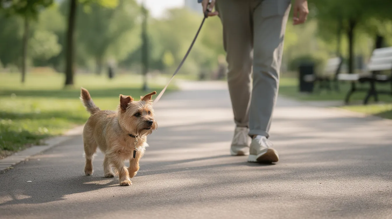
Most dogs cope well with cruciate surgery recovery, but owners need to plan realistically for a 3–4 month physical rehabilitation period. Rushing recovery is one of the most common causes of complications.
Approximate recovery phases:
|
Timeframe |
What to expect |
|---|---|
|
Weeks 0–2 |
Rest, wound care, very short toilet walks on a lead, ice packs if advised |
|
Weeks 3–6 |
Gradually increasing lead walks (5 minutes added per week), gentle range-of-motion exercises |
|
Weeks 7–12 |
Building endurance, introducing gentle hills, hydrotherapy if available |
|
Beyond 12 weeks |
Gradual return to near-normal activity once the vet confirms bone healing |
"Crate rest" or confinement in a UK home typically means using a secure room or large crate with non-slip flooring, blocking access to stairs with stairgates, preventing jumping onto or off furniture, and keeping the dog on a short lead for all outdoor time.
Veterinary physiotherapists and hydrotherapists (ideally ACPAT-registered practitioners) play a valuable role in the dog's recovery. Tailored physical therapy programmes help rebuild muscle mass, improve joint range of motion, and reduce complications. Hydrotherapy - swimming or underwater treadmill sessions - is particularly effective at building hindlimb strength without excessive joint loading.
Warning signs during recovery that need prompt veterinary review:
- Increased lameness or sudden worsening after a period of improvement
- Swelling, heat or discharge around the surgical wound
- Sudden yelping, exercise swelling, or reluctance to bear weight on the affected limb
- Any sign of the dog becoming unwell (off food, lethargic, feverish)
Long-term outlook, arthritis and mobility after cruciate disease
Cranial cruciate ligament disease is essentially an osteoarthritis-generating condition. Once the cruciate is damaged, some degree of arthritis in that knee is almost inevitable regardless of treatment. However, good management can keep many dogs comfortable and active for years.
Surgical stabilisation - particularly through osteotomy procedures - tends to slow arthritis progression and improve function compared with leaving the joint unstable. Conservative management can also achieve acceptable outcomes in well-selected cases, though arthritis typically advances more rapidly.
Realistic long-term expectations for most dogs include comfortable daily walks, moderate running and play. Some high-level working or agility dogs may have residual limitations depending on the severity of the original injury, the surgical technique used, and their age and conformation.
The pillars of long-term management are:
- Lifelong ideal body condition - keeping the dog lean reduces joint loading
- Appropriate daily exercise - consistent, moderate activity rather than sporadic intense sessions
- Environmental modifications - ramps, non-slip flooring, supportive bedding
- Nutritional support - including joint-supportive nutrients as part of an overall plan
- Veterinary monitoring - periodic check-ups allow pain scores, lameness patterns and muscle mass to be tracked, and medications or supplements adjusted as needed
Cruciate-related arthritis may flare in cold, damp weather or after unusually strenuous exercise. Learning to recognise these patterns allows owners to adjust activity and seek veterinary advice promptly when needed.
Will my dog injure the other knee?
This is one of the most common and understandably worrying questions owners ask after a cruciate ligament rupture.
The evidence is clear but sobering: studies suggest that around 40–60% of dogs that rupture one cranial cruciate ligament will eventually rupture the other knee. One UK study found bilateral rupture occurred in approximately 38.7% of affected dogs, with a mean interval of about 58 weeks (roughly 14 months), though this ranged from a few weeks to several years.
This high bilateral rate strongly supports the concept of an underlying cruciate ligament disease process affecting both joints rather than a one-off traumatic accident.
Practical steps that may reduce the risk or delay onset in the opposite knee:
- Strict weight control - keeping the dog at an ideal body condition score
- Progressive rehabilitation and muscle strengthening after the first surgery
- Avoiding uncontrolled high-impact exercise such as repetitive ball chasing and sudden skids
- Careful management of slippery floors at home
- Regular veterinary checks to monitor the other knee for early signs of instability or pain
Even if the second knee is eventually affected, many dogs cope well after staged surgeries and structured rehabilitation. Early detection often means a better outcome, so owners should remain vigilant for any change in gait or willingness to exercise.
Day-to-day management at home: flooring, stairs and lifestyle
Small daily changes at home can make a significant difference to dogs with cruciate disease, both during recovery and long-term.
Practical home modifications:
- Place non-slip rugs or runners on hard floors, particularly in hallways and where the dog turns corners
- Use ramps for getting in and out of the car or onto raised beds, rather than allowing jumping
- Install stairgates to limit unsupervised stair use, especially during the recovery period
- Block access to furniture that encourages jumping up or down
Once the vet approves, aim for controlled, low-impact exercise routines: short, frequent lead walks on even surfaces are far better than occasional long, intense outings. Consistency matters more than distance.
Avoid ball-launchers, sharp turns on slippery grass, and repetitive jumping in dogs with cruciate disease or a history of tear. For active dogs, swimming (once fully healed and with veterinary approval) is an excellent low-impact alternative.
During restricted activity periods, mental stimulation is essential to prevent frustration and boredom. Scent games, puzzle feeders, gentle training sessions and frozen enrichment toys can keep dogs content without stressing healing joints.
If considering knee braces or supports, work closely with your vet team. Fit, design, prescription and the overall management plan matter far more than marketing claims.
Weight management and nutrition for cruciate-affected dogs
Maintaining a lean body condition is one of the most powerful, evidence-supported actions owners can take to improve outcomes after a cruciate ligament injury. It is also one of the most commonly underestimated.
Every extra kilogram increases the force transmitted through the stifle with every step, worsening pain, accelerating arthritis, and placing additional strain on the uninjured knee. Overweight dogs are at substantially higher risk of cruciate rupture in the first place, and excess weight after surgery slows recovery and increases complication risk.
Practical weight management guidance aligned with WSAVA Global Nutrition Guidelines:
- Feed a complete and balanced diet appropriate for the dog's life stage
- Measure food portions accurately using scales rather than estimating
- Limit high-calorie treats - these should make up no more than 10% of daily intake
- Monitor body condition score regularly, ideally with veterinary input
- Consider therapeutic weight-management diets or veterinary nurse-led weight clinics for overweight dogs
Even modest weight loss - as little as 6–8% of body weight - can produce noticeable improvements in comfort and mobility in dogs with joint disease.
Alongside calorie management, joint-supportive nutrients can form part of an overall nutritional plan. Omega-3 fatty acids from fish oil or green-lipped mussel, for example, have evidence supporting their role in managing joint inflammation. For deeper reading on specific ingredients, the Alder & Hound guide to green-lipped mussel for dogs provides a thorough, evidence-informed overview.
Supplements are always an adjunct to, not a replacement for, weight loss and appropriate medical or surgical treatment.
Can joint supplements help dogs with cruciate ligament disease?
No supplement can repair a ruptured cranial cruciate ligament or replace the need for appropriate veterinary care. However, some ingredients may support joint health and help manage the secondary osteoarthritis that inevitably follows cruciate disease, when used as part of a comprehensive treatment plan.
Key ingredients commonly found in UK dog joint supplements:
- Green-lipped mussel - a natural source of omega-3 fatty acids and other bioactive lipids with evidence supporting anti-inflammatory effects in osteoarthritic joints
- Glucosamine - may support cartilage metabolism and synovial fluid quality, though evidence strength varies
- MSM (methylsulfonylmethane) - an organic sulphur compound with emerging data on supporting connective tissue health
- Boswellia serrata - a botanical extract with evidence for anti-inflammatory properties relevant to joint comfort
- Hyaluronic acid - a component of synovial fluid that may support joint lubrication; see the Alder & Hound guide to hyaluronic acid for dogs for details
- Long-chain omega-3 fatty acids (EPA/DHA) - well-studied for their role in modulating inflammatory pathways
Evidence strength varies between ingredients and between products. Response is individual - what works noticeably for one dog may be less apparent in another. High-quality formulations at meaningful inclusion levels are more likely to deliver benefit than generic low-dose blends.
Alder & Hound's ForeverRun™ is an example of a premium, evidence-informed joint supplement combining green-lipped mussel, MSM, glucosamine, boswellia extract, reishi mushroom, hyaluronic acid and vitamin C. It is designed to support joint health as part of an overall mobility plan - not to diagnose, treat, cure or prevent cruciate ligament rupture.
Always discuss supplement plans with your veterinary surgeon, especially if your dog is taking anti inflammatory medications or has underlying health conditions, to avoid interactions or unnecessary duplication.
Preventing cruciate ligament injuries where possible
Not all cruciate ligament ruptures are preventable. Genetics and conformation play a major role that owners cannot change. However, there are meaningful, evidence-supported steps that can reduce risk.
Key modifiable factors:
- Keep dogs lean - this is the single most impactful thing owners of at-risk breeds can do
- Avoid rapid weight gain - particularly in young large breed dogs during growth
- Build steady fitness - regular, consistent exercise rather than "weekend warrior" patterns
- Incorporate strength work - core and hindlimb strengthening exercises, ideally prescribed by a physiotherapist
- Avoid repetitive high-impact activities - constant ball chasing, leaping for frisbees, and sliding stops on hard or slippery surfaces are particularly risky in predisposed breeds
A study of agility dogs found that dogs engaged in regular core strength or conditioning programmes had lower odds of cruciate rupture, while increased body weight and spayed female status raised risk.
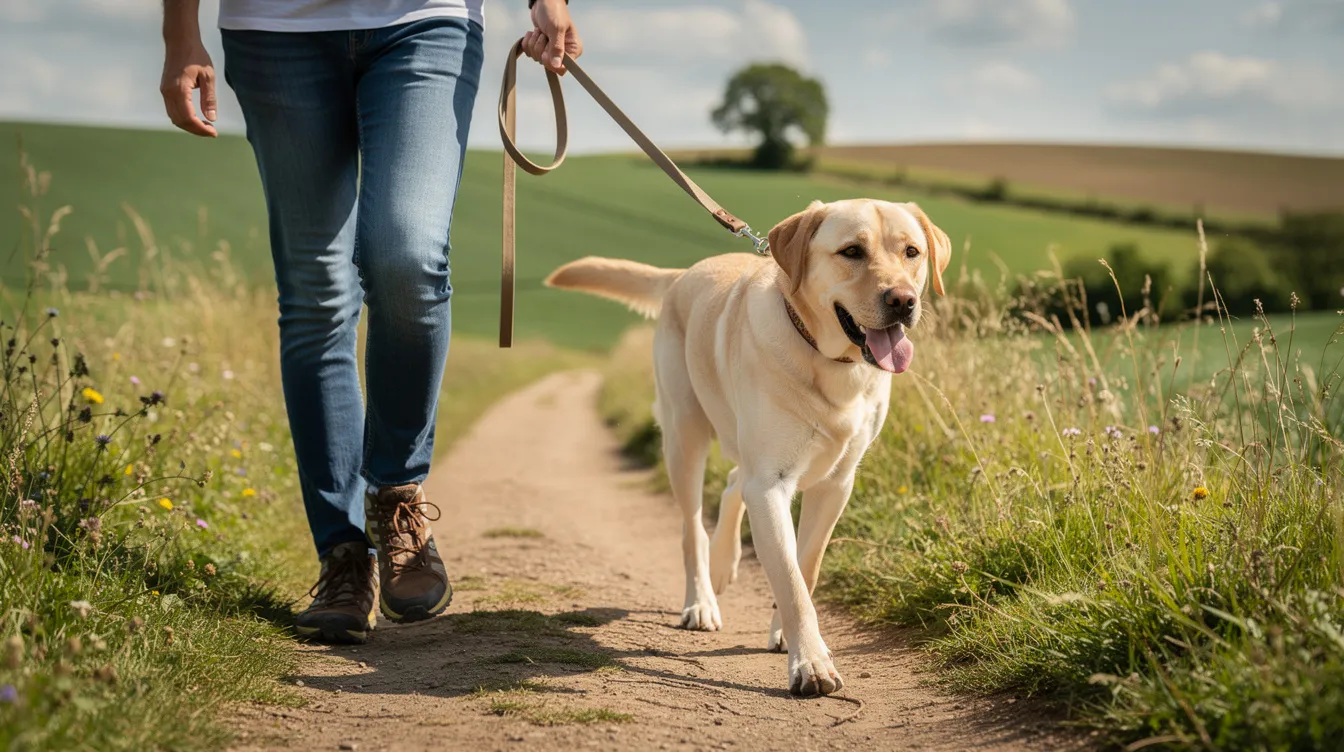
Practical protective exercise includes controlled lead walks, hill walking (once fit and healthy), swimming, and underwater treadmill hydrotherapy when appropriate. These build muscle around the stifle without the extreme forces generated by sudden twisting or jumping.
Responsible breeding practices may also help at a population level. Breeders who consider cruciate disease prevalence in their lines, alongside other health screening, can contribute to reducing risk across future generations.
Early veterinary assessment of any hindlimb lameness - even if mild or intermittent - gives the best chance of identifying developing cruciate disease and limiting further damage through early intervention.
Myth vs fact: common misconceptions about cruciate ligament injuries
Misinformation about cruciate disease is widespread online and can make decision-making harder for worried owners. Here are some of the most common myths, corrected with current evidence.
Myth: Cruciate injuries only happen after a big accident or trauma. Fact: The vast majority of canine cruciate ruptures result from gradual degenerative weakening of the ligament. The final rupture often occurs during normal, everyday activity.
Myth: Small dogs never need cruciate ligament surgery. Fact: While some small dogs can be managed conservatively, many benefit from surgery - particularly those with steep tibial plateau angles or high activity levels. Surgical treatment should be discussed on a case-by-case basis.
Myth: Surgery completely cures arthritis in the affected knee. Fact: Surgery stabilises the joint and reduces pain, but arthritis will still develop to some degree. The goal is to slow progression and maintain comfort, not to create a "brand new" knee.
Myth: Supplements can knit a torn ligament back together. Fact: No supplement repairs a ruptured ligament. Supplements may support joint health and help manage secondary osteoarthritis, but they are not a substitute for surgical repair or veterinary treatment.
Myth: If my dog is weight-bearing, the ligament can't be ruptured. Fact: Many dogs with a partially ruptured or even fully ruptured cranial cruciate ligament will still put some weight on the affected leg, especially after the initial acute phase. Weight-bearing does not rule out rupture.
Myth: Braces alone can reliably replace surgery in large dogs. Fact: While braces may have a limited role in some management plans, there is no robust evidence that bracing alone reliably replaces surgical intervention in large breed dogs with complete rupture.
Myth: Once one knee is fixed, the other will definitely be fine. Fact: Around 40–60% of dogs will eventually rupture the other knee. The underlying cruciate disease process often affects both joints.
Myth: My dog will never walk normally again after cruciate surgery. Fact: With appropriate surgical treatment and rehabilitation, most dogs return to comfortable daily walks and moderate activity. Complete return to high-level performance varies, but quality of life is typically very good.
Myth: Rest alone is enough for recovery. Fact: Structured physical rehabilitation - including controlled exercise, physiotherapy and, ideally, hydrotherapy - significantly improves outcomes compared with rest alone.
Myth: Only expensive referral surgeons can perform cruciate surgery. Fact: Many experienced primary-care veterinary surgeons perform extracapsular repairs, and some also offer osteotomy procedures. Referral to a specialist orthopaedic surgeon is advisable for complex cases but is not always necessary.
Related conditions that can look like cruciate disease
Not all hindlimb lameness or stiffness is caused by cranial cruciate ligament rupture. Incorrect self-diagnosis based on internet research can delay appropriate treatment, so veterinary assessment is always essential.
Important conditions that may mimic or coexist with cruciate disease:
- Hip dysplasia and hip osteoarthritis - common in similar breeds, causing hindlimb lameness and difficulty rising
- Luxating patella - the kneecap slipping out of its groove, causing intermittent skipping or hopping
- Lumbosacral disease - spinal nerve compression at the base of the spine, producing hindlimb weakness or pain
- Muscle or tendon injuries - strains, tears, or inflammation affecting the thigh or calf muscles
- Other knee problems - collateral ligament injury, isolated meniscal tears without cruciate rupture, or bone tumours
Some dogs, particularly larger breeds, may have multiple orthopaedic conditions simultaneously. A Labrador with both hip dysplasia and cruciate disease, for example, will have a more complex gait pattern and may need a staged treatment plan.
For deeper reading on related conditions, the Alder & Hound Complete Guide to Joint Health in Dogs covers arthritis, joint stiffness, and other mobility conditions in detail.
Only a veterinary examination - and often imaging - can reliably distinguish between these conditions and guide appropriate treatment.
Frequently asked questions about cruciate ligament rupture in dogs
How can I tell if my dog's lameness is serious? Any lameness lasting more than 24–48 hours, or any non-weight-bearing lameness, warrants veterinary assessment. Dogs that are getting worse rather than better, or that seem in significant pain, should be seen urgently.
Can a dog walk on a fully ruptured cruciate ligament? Yes, many dogs will begin to bear weight on the affected leg within a few days to weeks, even with a complete rupture. However, this does not mean the injury has healed - the joint remains unstable and arthritis will progress without appropriate management.
How long does recovery take after TPLO or TTA surgery? Most dogs are using the leg within days of surgery, but full recovery takes 3–4 months. Bone healing typically requires 8–12 weeks, during which exercise must be strictly controlled.
When can my dog go off the lead again? Most veterinary surgeons advise keeping dogs on a lead for at least 8–12 weeks post-surgery. Off-lead activity is usually only permitted once radiographs confirm bone healing and the vet is satisfied with recovery.
Are stairs allowed during recovery? Stairs should be avoided or very strictly limited during the first 8–12 weeks. Use stairgates and, if unavoidable, support the dog with a sling or harness on the lead.
How much does cruciate ligament surgery cost in the UK? Costs vary significantly. Extracapsular repairs at non-referral clinics may start from around £2,400. TPLO or TTA at referral hospitals typically ranges from £3,800 to £6,500 per knee depending on the dog's size and the clinic. Always request a detailed written estimate.
Does pet insurance cover cruciate surgery? Most comprehensive lifetime policies cover cruciate ligament surgery, but pre-existing conditions, policy limits and excesses vary. Check your policy carefully and inform your insurer promptly.
What is the success rate for cruciate surgery? Success rates are generally high. Studies report good or excellent outcomes in 85–95% of surgically treated dogs, though "success" is usually defined as comfortable mobility rather than a perfectly normal joint.
What happens if I can't afford surgery? Discuss this honestly with your vet. Conservative management is an option, particularly for smaller dogs. Payment plans, charity assistance, and choosing a less costly surgical technique may also be possibilities.
Is my older dog too old for surgery? Age alone is not a contraindication. Many older dogs undergo cruciate surgery successfully. The decision depends on overall health, anaesthetic risk, and the dog's quality of life. Your vet can help assess this.
Is hydrotherapy essential for recovery? It is not essential, but it is highly beneficial. Hydrotherapy helps rebuild muscle mass with reduced joint stress and can speed recovery. If unavailable, structured land-based exercises prescribed by a physiotherapist are a good alternative.
When should I start physiotherapy? Many veterinary physiotherapists see dogs within the first 1–2 weeks after surgery for gentle range-of-motion work. Active strengthening programmes typically begin from around week 3–4 onwards, guided by the healing timeline.
Will my dog need medication long-term? Some dogs need ongoing pain relief or anti-inflammatory medication. Others manage well with weight control, exercise management and nutritional support alone. Your vet will tailor a long-term plan to your dog's needs.
Should I get a second opinion or ask for a referral? If you have any uncertainty about the diagnosis or recommended treatment, requesting a referral to a veterinary orthopaedic specialist is entirely reasonable and most vets will support this decision.
Can a partially ruptured cruciate ligament heal on its own? Unfortunately, the cranial cruciate ligament has very poor healing capacity. Partial tears usually progress to complete rupture over time, particularly in larger or more active dogs. Early intervention often produces better outcomes.
Alder & Hound's role: education, joint support and ongoing care
Alder & Hound is a UK-based educational resource dedicated to canine mobility and healthy ageing. Our goal is to help owners understand joint health, make informed decisions alongside their veterinary team, and provide the best possible long-term support for their dogs.
The Alder & Hound Canine Mobility Knowledge Centre offers in-depth guides on joint health, arthritis, specific ingredients and condition-focused articles that complement veterinary advice. Whether you are researching green-lipped mussel, boswellia, glucosamine, hyaluronic acid or MSM, our evidence-informed ingredient library provides balanced, referenced information to help you evaluate your options.
A premium joint supplement such as ForeverRun™ can form part of a broader long-term management plan after cruciate surgery or conservative care - alongside weight control, appropriate exercise, environmental modifications and, where necessary, prescribed medications. It is not a replacement for veterinary treatment, and no supplement from Alder & Hound or any other company can diagnose, treat, cure or prevent cruciate ligament rupture.
We encourage you to explore our cornerstone content, including the Complete Guide to Joint Health in Dogs and condition-specific guides on arthritis and joint stiffness, to deepen your understanding of canine mobility at every life stage.
If you would like to receive ongoing educational updates, mobility-focused content and practical advice for caring for your dog's joints, we invite you to sign up through our website.
Key takeaways for owners of dogs with cruciate ligament injuries
Cranial cruciate ligament disease is one of the most common causes of hindlimb lameness in UK dogs. It is usually a degenerative condition - the ligament weakens over time before eventually failing - rather than a purely traumatic event.
Early veterinary assessment is essential. Whether your vet recommends surgical treatment or conservative management, timely diagnosis and an appropriate treatment plan dramatically improve most dogs' comfort and long-term mobility.
Keeping your dog lean, fit and well-muscled is one of the most powerful things you can do at every stage: before injury, during recovery, and for the rest of their life. Simple home adjustments - non-slip flooring, ramps, stairgates - protect healing knees and improve daily comfort.
Arthritis is almost inevitable after cruciate rupture, but it does not have to mean a poor quality of life. Long-term, proactive management - including nutrition, appropriate exercise, environmental changes, medication where needed, and evidence-informed supplements - can maintain comfort and mobility for years.
Owners should remain aware that the other knee may also be affected in time, and ongoing monitoring, weight control and muscle conditioning are the best defences.
You are not alone in navigating this. Your veterinary team, qualified rehabilitation professionals, and educational resources like the Alder & Hound Canine Mobility Knowledge Centre are all here to support you and your dog through every stage - from diagnosis through recovery and beyond.