

Elbow Dysplasia in Dogs: Complete Evidence‑Informed Guide for UK Owners
Elbow dysplasia in dogs is a group of developmental elbow joint problems and one of the most common causes of forelimb lameness in the United Kingdom, often affecting medium to large breeds and leading to early arthritis if it is not managed well. If your dog has recently been diagnosed, or if you have noticed your puppy or young adult limping on a front leg, this guide will help you understand the different types of elbow dysplasia, what causes it, the signs to look for, how vets diagnose it, and the treatment and long-term management options that can protect your dog's comfort and mobility for years to come.
Written by the team at Alder & Hound, this resource is for UK dog owners who want clear, evidence-informed advice on canine elbow joint health, including pain relief, surgery and non-surgical care, prevention, working with your vet, and where joint supplements may fit into an overall support plan. It draws on current veterinary evidence, UK screening data and real-world clinical insights to give you the information you need - clearly and without unnecessary alarm.
Quick answer: what is elbow dysplasia in dogs?
Elbow dysplasia is a group of developmental conditions affecting the canine elbow joint - the complex joint where the humerus, radius and ulna meet. It is the most common cause of forelimb lameness in dogs, particularly in medium, large and giant breed dogs, and a leading cause of early-onset arthritis in the affected elbow.
The term canine elbow dysplasia covers four distinct abnormalities: fragmented medial coronoid process (FCP), ununited anconeal process (UAP), osteochondritis dissecans OCD of the humeral condyle, and elbow incongruity - a poor "fit" between the joint surfaces. These conditions can occur alone or in combination.
Elbow dysplasia itself cannot be reversed. However, many dogs live active, comfortable lives with the right combination of weight control, controlled exercise, pain relief, physiotherapy and, where appropriate, surgical intervention. Early diagnosis and a tailored treatment plan can significantly reduce the impact of secondary osteoarthritis and improve long-term quality of life.
Elbow dysplasia can lead to secondary osteoarthritis in dogs, which is why prompt, evidence-informed management matters so much.
At Alder & Hound, we are a UK canine joint-health specialist dedicated to providing trusted information and premium joint supplements - including ForeverRun™ - to support long-term mobility alongside veterinary care.
Key points:
- Elbow dysplasia is a developmental condition, not a single disease
- It most commonly affects medium to large breeds during growth
- Four distinct abnormalities are associated with elbow dysplasia
- Early diagnosis and treatment improve long-term outcomes
- Lifelong management is usually required, but most dogs respond well
When should you contact your vet about elbow lameness?
Any persistent front leg lameness in a puppy or young adult dog should be assessed by a vet within a few days, even if your dog is still willing to exercise. Dogs are remarkably good at masking discomfort, and early assessment opens the door to earlier, more effective treatment.
Red-flag signs - same day veterinary attention:
- Inability to bear weight on a front leg
- Marked elbow swelling, heat or obvious deformity
- Severe pain, yelping or crying on movement
- Sudden worsening of existing lameness
- Collapse, lethargy or signs of feeling unwell
Non-emergency but prompt signs - within a week:
- Intermittent limping on one or both front legs
- Stiffness when rising after rest
- Bunny-hopping gait or shortened stride in the front limbs
- Reluctance to jump, play or complete usual walks
- Consistently turning one front paw outwards
Because elbow dysplasia is often bilateral - a large UK study found both elbows affected in around 61% of cases - lameness can look surprisingly subtle. Both front legs may be equally stiff, so there is no obvious head-nod or limp, just a general reluctance to move freely.
Delaying assessment allows more cartilage damage to accumulate and brings forward the onset of elbow osteoarthritis. Do not rely on rest alone.
Important: Never give your dog human painkillers such as ibuprofen or paracetamol without veterinary direction. Many human medications are toxic to dogs.
How the normal canine elbow joint works
Understanding what a healthy elbow looks like helps explain what goes wrong in elbow dysplasia.
The canine elbow is a complex joint formed by three bones: the humerus (upper arm), the radius and the ulna (the two bones of the forearm). These three bones must fit together precisely. When they do, forces are distributed evenly across the joint surface during walking, running, jumping and play.
Several key structures are especially relevant to elbow dysplasia:
- Medial coronoid process - a small, load-bearing projection on the inner (medial) side of the ulna. It sits beneath the medial humeral condyle and bears significant weight during movement.
- Anconeal process - a bony hook at the top of the ulna that locks into the humerus to stabilise the elbow during extension.
- Trochlear notch - the curved surface of the ulna that cradles the humerus, allowing smooth flexion and extension.
- Articular cartilage - the smooth, low-friction surface covering all bone ends inside the joint. Joint fluid, which contains hyaluronic acid, lubricates and nourishes this cartilage.
The concept of joint congruity is central. A well-matched fit between the joint surfaces distributes pressure evenly. Even small mismatches - perhaps only a millimetre or two - can dramatically increase local pressure on one area, particularly the medial aspect of the joint. This is why even subtle joint incongruity can trigger significant problems over time.
Graphic placeholder: Normal elbow vs elbow dysplasia anatomy - labelled diagram comparing a healthy elbow joint with an affected elbow joint, highlighting the medial coronoid process, anconeal process, trochlear notch and humeral condyle.
What is elbow dysplasia? (umbrella term explained)
The word "dysplasia" comes from the Greek for abnormal development. Elbow dysplasia literally means abnormal growth of the elbow. Crucially, it is not a single disease but an umbrella term covering several related developmental conditions that affect how the elbow joint forms during growth.
The four main components are:
- Fragmented medial coronoid process (FCP), also called medial coronoid disease
- Ununited anconeal process UAP
- Osteochondritis dissecans (OCD) of the medial humeral condyle
- Elbow incongruity - step, notch or shape mismatch between the radius, ulna and humerus
Different specialist centres sometimes use slightly different terminology. For example, you may hear "medial compartment disease" when the damage is concentrated on the inner side of the joint, or "medial coronoid disease" instead of "fragmented coronoid process." These terms describe similar underlying problems.
All four abnormalities share a common outcome: they create uneven loading within the elbow joint, damage the articular cartilage, and ultimately drive elbow osteoarthritis if not managed well. The disease process usually begins while the skeleton is growing - mainly up to around 10–12 months of age - but clinical signs may not become obvious until later.
How does elbow dysplasia compare with hip dysplasia?
If you are already familiar with hip dysplasia in dogs, the parallels are clear. Both involve abnormal joint shape during development, both lead to instability or incongruity, and both progress to secondary osteoarthritis. The key difference is anatomy: the elbow is a far more tightly constrained joint than the hip, which means even small developmental abnormalities can have a significant impact.
The four main forms of elbow dysplasia: overview
The following table summarises the four main forms of elbow dysplasia at a glance.
|
Condition |
Affected structure |
Typical age at diagnosis |
Commonly affected breeds |
Key features |
|---|---|---|---|---|
|
Fragmented coronoid process (FCP) |
Medial coronoid process (ulna) |
5–12 months |
Labrador Retrievers, Rottweilers, Bernese Mountain Dogs, Golden Retrievers |
Most common form; subtle on X-ray; CT or arthroscopy often needed |
|
Ununited anconeal process (UAP) |
Anconeal process (ulna) |
4–8 months |
German Shepherd Dogs, Basset Hounds, Bernese Mountain Dogs |
Failure of bone fusion; visible on flexed X-ray |
|
Osteochondritis dissecans (OCD) |
Medial humeral condyle |
4–10 months |
Large and giant breeds; males more often |
Cartilage flap or defect; good surgical outcomes if treated early |
|
Elbow incongruity |
Radius–ulna length mismatch or notch shape |
Variable |
Any predisposed breed |
Often co-exists with FCP; CT essential for accurate assessment |
Many dogs have more than one lesion in the same joint. CT imaging studies have shown that medial coronoid disease combined with elbow incongruity is particularly common, occurring in over half of affected elbows in some cohorts.
All four forms produce similar outward signs - front leg lameness, stiffness, pain on elbow manipulation - so diagnostic imaging is essential to tell them apart and guide treatment choices.
Graphic placeholder: The four forms of elbow dysplasia comparison - side-by-side infographic summarising each condition.
Fragmented medial coronoid process (FCP / medial coronoid disease)
Fragmented medial coronoid process is the most frequently diagnosed form of elbow dysplasia. It occurs when the medial coronoid process - that small, weight-bearing projection on the inner side of the ulna - develops small cracks, fissures or fragments due to abnormal mechanical loading and micro-fractures.
Medial coronoid disease affects large-breed dogs like Labrador Retrievers, Bernese Mountain Dogs, Rottweilers, Golden Retrievers and Newfoundlands. Fragmented coronoid process is a common issue in elbow dysplasia overall. Most dogs are diagnosed between 5 and 12 months of age, although some are not recognised until middle age when secondary arthritis becomes apparent. In one CT imaging cohort, approximately 69% of dogs were diagnosed at two years of age or younger.
Common signs include:
- Intermittent forelimb lameness, often worse after exercise
- Stiffness after rest
- Pain on elbow flexion, extension or rotation
- Reduced range of motion
- Muscle loss over the affected shoulder
Diagnosis: Standard radiographs often struggle to visualise the coronoid process clearly. A CT scan is particularly useful for detecting subtle lesions, including hairline fractures and subchondral bone changes. Arthroscopy allows direct visualisation of the affected joint surface and is considered the gold standard for confirming the diagnosis and assessing cartilage damage.
Treatment options:
- Conservative management - weight control, controlled exercise, NSAIDs, physiotherapy, hydrotherapy and joint supplements - may be appropriate for mild disease or older dogs
- Arthroscopic surgery - fragment removal can lead to rapid clinical improvement in many cases
- Subtotal coronoid ostectomy (SCO) - used for diffuse stress fracturing where no single fragment can be removed
- Proximal ulnar osteotomy or proximal abducting ulnar osteotomy (PAUL) - when significant elbow incongruity contributes to coronoid overload
Prognosis: Many dogs improve significantly with treatment, but some degree of elbow osteoarthritis is expected long-term. Outcome depends on age at treatment, severity of cartilage damage and the quality of ongoing management.
Ununited anconeal process (UAP)
The anconeal process is a bony prominence at the top of the ulna that normally fuses with the rest of the bone by around five to six months of age. An ununited anconeal process occurs when this fusion fails, leaving the anconeal process loose or partially attached. This creates instability at the back of the elbow joint.
Suspected causes include osteochondrosis, mismatched growth rates between the radius and ulna (a relatively short radius pushing the anconeal process out of alignment), and genetic predisposition.
Breeds especially prone include German Shepherd Dogs, Basset Hounds and Bernese Mountain Dogs. UAP typically presents between four and eight months of age and is often bilateral.
Signs to watch for:
- Weight-bearing but obvious lameness
- Elbow joint effusion (swelling)
- Reduced range of motion, crepitus (clicking)
- Pain on full flexion or extension
- Muscle wasting if the condition is chronic
Diagnosis: A flexed mediolateral radiograph often reveals the unfused anconeal process clearly. CT may be used to assess concurrent lesions and elbow incongruity.
Treatment: In young dogs, early surgical reattachment of the anconeal process with a screw can be successful. When diagnosis is delayed, removal of the fragment combined with procedures such as proximal ulnar osteotomy to relieve abnormal forces may be needed. Lifelong management of secondary osteoarthritis is typically required.
Prognosis: Better when surgery is performed before severe cartilage damage, in younger dogs. Many dogs achieve good comfort and function with a combination of surgical and medical care, though arthritis risk remains.
Osteochondritis dissecans (OCD) of the elbow
Osteochondrosis is a developmental problem where the normal conversion of cartilage to bone (endochondral ossification) does not proceed properly in rapidly growing dogs. Osteochondrosis typically affects young, rapidly growing large breeds.
In the elbow, OCD specifically refers to a flap or defect of thickened, diseased cartilage - usually on the medial humeral condyle - that may partially detach or float within the joint, causing pain and inflammation. This cartilage flap acts as a mechanical irritant and accelerates the disease process within the joint.
Risk factors include:
- Fast growth in large and giant breed dogs
- Genetic predisposition
- Excessive energy or calcium intake during puppyhood
- Trauma or over-exercise during the growth period
- Male dogs may be more commonly affected
Signs: Forelimb lameness usually appears before one year of age, often accompanied by joint pain, stiffness, reduced willingness to exercise and sometimes low mood due to chronic discomfort.
Diagnosis: Clinical examination and orthopaedic testing, followed by radiographs under sedation to assess the humeral condyle. Advanced imaging techniques include CT and MRI for elbow dysplasia cases where multiple lesion types may co-exist.
Treatment: Arthroscopic surgery to remove the OCD lesion - debridement and removal of the cartilage flap - is the standard approach. For larger defects, resurfacing techniques such as osteochondral autograft transfer or synthetic osteochondral replacement may be considered at specialist centres.
Prognosis: Most dogs experience significant improvement after surgery. However, osteoarthritis begins as soon as OCD develops, so long-term joint-supportive management remains essential.
Elbow incongruity and abnormal joint shape
Elbow incongruity means a mismatch in shape or length between the radius, ulna and humerus. Even small differences can lead to abnormal load distribution in joints, concentrating forces on the medial compartment and accelerating cartilage damage.
Main types of incongruity:
- Short radius - the radius is slightly shorter than the ulna, shifting more load onto the coronoid process
- Short ulna - the ulna is relatively short, pushing the anconeal process into the humerus
- Trochlear notch dysplasia - the curved cradle of the ulna is abnormally shaped, creating a poor fit
- Radio-ulnar conflict - the radial head presses against the coronoid process under load
Incongruity can be primary (due to abnormal growth patterns) or secondary (following trauma or other developmental conditions). It is a major driver of medial compartment overload and is frequently found alongside medial coronoid disease.
Diagnosis: CT imaging is usually essential. Studies comparing X-ray and CT have shown that CT detects incongruity in significantly more dogs than radiographs alone. In healthy dogs, the radioulnar "step" measures approximately 0.85 mm, while in affected dogs it averages around 2.33 mm on CT - a seemingly small difference with large clinical consequences.
Treatment approaches:
- Conservative management may be chosen for mild cases or when advanced arthritis is already present
- Corrective osteotomy - a controlled bone cut - can improve joint congruity in young dogs. Procedures include proximal ulnar osteotomy (PUO), proximal dynamic ulnar osteotomy and proximal abducting ulnar osteotomy (PAUL)
- Load-altering surgery such as sliding humeral osteotomy (SHO) aims to shift weight away from the damaged medial compartment
An "osteotomy" simply means a planned surgical cut in a bone to realign it or change how forces travel through the joint. "Load shifting" refers to redirecting pressure away from the most damaged area of the joint surface.
Prognosis: The aim is to reduce joint pain and slow progression of osteoarthritis, not to create a "perfect" elbow. Outcomes depend on age at surgery and existing cartilage damage.
Why does elbow dysplasia develop? (causes and risk factors)
Elbow dysplasia is a multifactorial disease, meaning no single factor causes it. Rather, a combination of genetics, growth patterns and environmental influences during the critical skeletal development period determines whether a dog develops the condition.
Genetics
Genetic predisposition plays a major role in elbow dysplasia development. Heritability estimates range from 0.10 to 0.38 depending on breed and lesion type, with some studies reporting even higher values - for example, approximately 0.57 for medial coronoid process disease in German Shepherd Dogs. More than 100 genes are associated with elbow dysplasia, reflecting the complexity of the genetic architecture. Dogs that appear clinically normal may still carry and pass on genetic risk.
Growth rate and body weight
Rapid growth and excess body weight increase mechanical load on the developing elbow joint. In a landmark study of Labrador Retrievers, dogs fed ad libitum (unrestricted food) had approximately 26% higher body mass and developed significantly more radiographic osteoarthritic change by age six compared with calorie-restricted dogs.
Nutrition
A complete, balanced puppy diet appropriate for breed size is essential. The WSAVA Global Nutrition Guidelines emphasise avoiding oversupplementation of calcium, vitamin D or energy in growing large-breed puppies, as this can disrupt normal skeletal development.
Exercise and environment
Repetitive high-impact activities - long sessions of ball-chasing, jumping from height, frequent stairs - may worsen micro-trauma in genetically susceptible elbows. Exercise is unlikely to be the sole cause, but it can accelerate the abnormal development in predisposed puppies.
Other factors
Early neutering is an area of ongoing research. Some studies suggest it may influence growth plate closure timing and body composition, but the evidence specifically for elbow dysplasia is not yet conclusive. Trauma can unmask an underlying predisposition rather than create the condition from scratch.
Responsible breeding and sensible growth management can reduce risk but cannot guarantee prevention.
Which dogs are most at risk? (breeds and age)
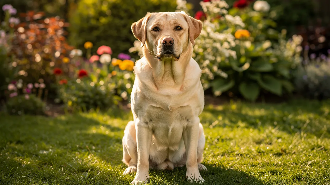
Elbow dysplasia affects large and giant breed dogs most frequently, though it can occur in medium-sized breeds and occasionally in crossbreeds. It should not be ruled out based on size alone.
|
Breed |
Main lesions seen |
Typical age at diagnosis |
Notes |
|---|---|---|---|
|
Labrador Retrievers |
FCP, OCD, incongruity |
5–18 months |
Very commonly affected; bilateral disease frequent |
|
Golden Retrievers |
FCP, OCD |
5–18 months |
Elbow scoring recommended |
|
German Shepherd Dogs |
UAP, FCP |
4–12 months |
High heritability for coronoid disease |
|
Bernese Mountain Dogs |
FCP, UAP, OCD |
5–12 months |
Multiple lesion types common |
|
Rottweiler |
FCP, incongruity |
5–18 months |
Bilateral disease common |
|
Newfoundland |
FCP, OCD |
6–18 months |
Giant breed; weight management critical |
|
English Springer Spaniel |
FCP |
5–18 months |
Medium breed; often overlooked |
|
Basset Hound |
UAP |
4–8 months |
Chondrodystrophic breed |
UK screening data from the BVA/KC elbow grading scheme shows that while a large majority of screened dogs achieve Grade 0 (no radiographic changes), a notable minority of large breeds show evidence of disease. For example, among Saint Bernards, approximately 20% had Grade 1–3 changes.
Symptoms of elbow dysplasia often appear between 5 and 18 months of age, though some dogs only present when secondary osteoarthritis becomes advanced - sometimes at four to six years or older. Symptoms can appear as early as four months in some individuals.
Elbow dysplasia is often bilateral, affecting both elbows in up to 60–80% of cases. If your dog belongs to a predisposed breed, discuss early screening and elbow scoring with your vet and breeder.
Graphic placeholder: Breeds most at risk infographic - summarising top predisposed breeds and age of onset.
Signs and symptoms of elbow dysplasia in dogs
Early signs you might notice at home:
- Intermittent front leg limping, sometimes swapping sides
- Stiffness when getting up after rest
- Decreased enthusiasm for walks or lagging behind
- Licking or chewing at the elbow area
- Difficulty jumping into the car or onto furniture
- Turning one front foot outward when standing
Common symptoms include lameness and stiffness after rest. Lameness often worsens after exercise or long walks and may seem to improve with rest, leading owners to assume a simple sprain.
Gait changes to watch for:
- Head nodding - the head lifts when the painful leg bears weight
- Shortened stride in the affected limb
- Elbows held slightly away from the body
- Shifting weight backwards onto the hindlimbs
Later-stage signs:
- Muscle loss over the shoulders
- Visible thickening around the affected elbow joints
- Audible clicking or crepitus
- Reluctance to be handled or irritability, often due to chronic joint pain
- Behavioural changes such as reduced playfulness or withdrawal
Bilateral elbow dysplasia can obscure lameness appearance in dogs. When both elbows are equally sore, there may be no obvious limp - just a general unwillingness to exercise or a stiff, shuffling gait.
What your vet may notice on examination:
- Pain on flexion, extension or pronation/supination of the elbow
- Reduced range of motion
- Joint effusion (fluid around the joint)
- Muscle asymmetry between front limbs
Graphic placeholder: Early warning signs checklist - a downloadable/printable resource for owners.
How vets diagnose elbow dysplasia
Diagnosis of elbow dysplasia typically involves physical examination and imaging, progressing from simpler to more advanced techniques as needed.
Clinical examination
Your vet will begin with a thorough history and physical examination. This includes watching your dog walk and trot (gait assessment), palpating the elbow joint for pain, swelling or heat, and testing range of motion. Specific manipulation tests can stress the medial compartment and anconeal process to localise pain.
Radiography (X-rays)
Radiography is commonly used to diagnose elbow dysplasia and is available at most first-opinion practices. Sedation or light anaesthesia is usually needed for high-quality images. X-rays can reveal an ununited anconeal process, osteophytes (new bone), sclerosis and some forms of incongruity. However, they have significant limitations for detecting subtle coronoid disease or early cartilage damage.
CT scan
CT scans provide detailed images of elbow joint abnormalities, generating three-dimensional cross-sectional views that look beneath the cartilage. CT is particularly useful for detecting subtle lesions in dogs, including hairline coronoid fractures, subchondral bone defects and precise measurement of joint incongruity. It is typically available at veterinary referral centres.
Arthroscopy
Arthroscopy is a minimally invasive surgery using a tiny camera inserted into the joint through a small incision. It allows direct visualisation of the elbow joint, confirming cartilage damage, assessing severity and enabling treatment (such as fragment removal) in the same procedure.
Other diagnostic tools
Advanced diagnostic imaging may include MRI for soft tissue assessment, though this is less commonly used for primary elbow dysplasia. Blood tests may be performed to assess fitness for anaesthesia or rule out other causes of lameness.
Graphic placeholder: Diagnostic pathway flowchart - from first vet visit through clinical examination, X-rays, CT and arthroscopy.
Non-surgical management: everyday treatment for elbow dysplasia
Not all dogs with elbow dysplasia require surgery. Many dogs are managed successfully - either instead of surgery or alongside it - with a combination of lifestyle changes, medication and supportive therapies. Non-surgical treatments include weight management and physiotherapy as core pillars. Long-term management often requires multimodal approaches combining several strategies.
Weight management
Keeping your dog lean is the single most important modifiable factor. Every extra kilogram of body weight increases load across the affected elbow. Evidence from longitudinal studies consistently shows that maintaining a healthy body condition slows osteoarthritis progression.
Controlled exercise
Regular, low-impact activity maintains muscle mass and joint mobility without overloading the elbow. Aim for frequent, shorter walks on even surfaces. Avoid sharp turns, ball throwing on slippery ground, repetitive jumping and prolonged high-speed running.
Physiotherapy
A qualified canine physiotherapist can design an individualised programme including range-of-motion exercises, strengthening of the shoulder and core muscles, and exercises you can do at home between sessions. Physical therapy helps maintain function and can reduce the need for higher doses of pain medication.
Hydrotherapy
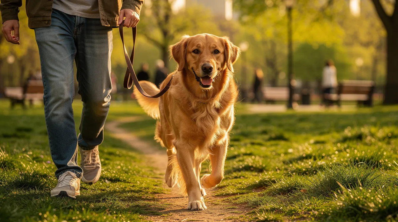
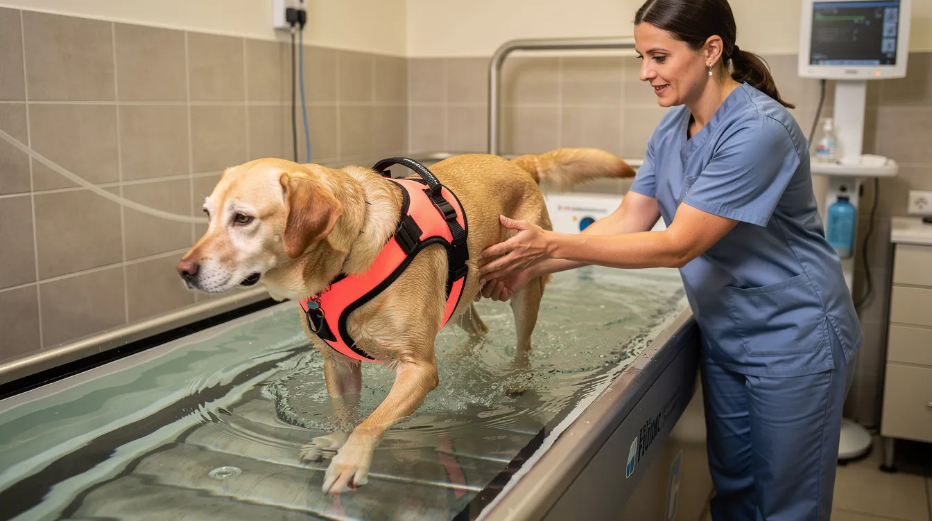
Underwater treadmill work or controlled swimming improves muscle strength and cardiovascular fitness with reduced joint impact. Your vet can refer you to a reputable centre.
Environmental modifications
- Lay non-slip rugs or runners over hard flooring
- Use ramps instead of stairs or for car access
- Provide supportive orthopaedic bedding in a warm, draught-free location
- Keep nails trimmed short to improve traction
Do and avoid list:
|
Do |
Avoid |
|---|---|
|
Multiple short, steady lead walks daily |
Long off-lead romps on uneven ground |
|
Low-impact activities like scentwork |
Repetitive ball throwing or fetch |
|
Regular weight checks |
Allowing excess weight gain |
|
Physiotherapy exercises at home |
Jumping in/out of cars without a ramp |
|
Regular veterinary reviews |
Ignoring subtle changes in mobility |
Graphic placeholder: Daily management checklist - core lifestyle strategies.
Pain relief and medical therapies for elbow dysplasia
Effective pain management for elbow dysplasia uses a multimodal approach - combining different classes of medication and non-drug therapies to improve comfort while minimising side effects.
NSAIDs (non-steroidal anti-inflammatory drugs)
NSAIDs remain the mainstay for managing inflammation and joint pain in dogs affected by elbow osteoarthritis. They must be prescribed by a vet and monitored with periodic blood tests, particularly liver and kidney function checks. Common veterinary NSAIDs include meloxicam, carprofen and robenacoxib.
Adjunctive medications
Where NSAIDs alone are insufficient, veterinary surgeons may add:
- Paracetamol (dogs only, under strict veterinary direction)
- Gabapentin or amantadine for chronic pain modulation
- Short courses of opioids during severe flare-ups or post-operatively
Intra-articular treatments
Some vets or specialists may offer injections directly into the affected joint:
- Hyaluronic acid injections can supplement the joint's own lubrication
- Corticosteroid injections may provide short-term relief in specific situations
Both have potential benefits and limitations; discuss realistic expectations with your specialist.
Regenerative medicine
Platelet rich plasma (PRP) and stem cell therapy are emerging areas with promising but still developing evidence. Stem cells and PRP aim to reduce inflammation and potentially support tissue healing, but protocols and outcomes vary considerably. These treatments are typically offered at specialist centres and can be costly. Owners should discuss expectations openly with their specialist.
Never give human painkillers such as ibuprofen, aspirin or naproxen to your dog. These can cause serious, sometimes fatal, side effects.
Joint supplements and nutritional support
Joint supplements play a supporting role in the long-term management of dogs with elbow dysplasia and secondary osteoarthritis. They are not cures for the underlying developmental abnormality, but they can contribute to ongoing joint comfort and function as part of a broader management plan.
Key ingredients used in UK canine joint supplements:
- Green Lipped Mussel - a natural source of omega-3 fatty acids and glycosaminoglycans; evidence for supporting joint comfort in osteoarthritic dogs is relatively strong. Read our full Green Lipped Mussel for Dogs guide.
- Glucosamine and chondroitin - widely used to support cartilage health; evidence is mixed but generally supportive of modest benefit in combination products
- MSM (methylsulfonylmethane) - a source of organic sulphur; may support joint comfort and reduce inflammation
- Omega-3 fatty acids - well-evidenced for anti-inflammatory effects in osteoarthritis
- Boswellia serrata - a botanical extract with anti-inflammatory properties; emerging evidence supports its use in canine joint support. See our Boswellia for Dogs guide.
- Hyaluronic acid - a component of synovial fluid and cartilage; oral supplementation may support joint lubrication. Learn more in our Hyaluronic Acid for Dogs guide.
-
Vitamin C - an antioxidant that supports collagen synthesis
Alder & Hound's ForeverRun™ is a premium, multi-ingredient formulation combining Green Lipped Mussel, MSM, glucosamine, Boswellia extract, Reishi mushroom, hyaluronic acid and vitamin C. A product like this can fit naturally into a broader management plan when discussed with your vet.
Before adding any supplement, ensure your dog is eating a complete, balanced core diet - especially important during the growth phase for large-breed puppies. Allow six to eight weeks to assess benefit, avoid stacking multiple similar products, and watch for any gastrointestinal upset.
What joint supplements can and cannot do:
-
✔ Support ongoing joint comfort as part of a multimodal plan
-
✔ Provide building blocks for cartilage and joint fluid maintenance
-
✔ Help manage secondary osteoarthritis alongside veterinary treatment
-
✗ Cannot correct the underlying developmental abnormality
-
✗ Cannot reverse existing cartilage loss
-
✗ Should not replace veterinary assessment or prescribed medication
For a comprehensive overview, visit our Complete Guide to Joint Health in Dogs.
Surgical options for elbow dysplasia
Surgical treatment may be recommended based on lesion type, age, severity of elbow incongruity and degree of cartilage damage. However, surgery is not mandatory for every dog with elbow dysplasia. Surgical treatments aim to minimise osteoarthritis progression, reduce pain and improve function - but they cannot reverse existing cartilage damage or guarantee a "normal" joint.
Arthroscopy and fragment removal
Arthroscopic surgery is the most commonly performed procedure for FCP and OCD lesions. Through small incisions, a camera and specialised instruments allow the surgeon to remove loose fragments, debride diseased cartilage and assess the joint surface. Both elbows can sometimes be treated in a single anaesthetic. Arthroscopic fragment removal can lead to rapid clinical improvement, with most dogs bearing weight within days. Around 85% of dogs show improvement in lameness after treatment, though the degree of improvement varies.
Corrective osteotomies and load-altering surgery
When elbow incongruity is a significant factor, surgical procedures that realign or redirect forces through the joint may be recommended:
- Proximal ulnar osteotomy (PUO) - a cut in the ulna that allows the joint surfaces to realign
- Proximal abducting ulnar osteotomy (PAUL) - shifts the ulna outward to offload the damaged medial compartment
- Proximal dynamic ulnar osteotomy - a variant allowing gradual self-correction
- Sliding humeral osteotomy (SHO) - redirects forces from the medial to the lateral side of the joint
These procedures are technically demanding, typically performed at referral centres, and require longer recovery periods with careful rehabilitation.
Salvage procedures
For dogs with severe OA or advanced arthritis where other treatments have failed:
- Unicompartmental elbow resurfacing (CUE) - replaces the damaged medial compartment surfaces
- Total elbow replacement - a salvage procedure for severe cases where complete joint replacement is performed
Total elbow replacement and elbow replacement procedures generally are specialised, high-cost and available at only a small number of UK referral centres. Outcomes can be variable, and careful patient selection is essential.
Factors influencing surgical management decisions:
- Age and general health of the dog
- Severity of changes on CT or arthroscopy
- Activity level and owner expectations
- Financial considerations and access to specialist care
Graphic placeholder: Surgical options comparison table - purpose, lesion types, typical candidates, recovery time and long-term goals.
Rehabilitation and recovery after elbow surgery
Recovery after elbow surgery is a gradual process. Whether your dog has had arthroscopic surgery, fragment removal, an ulnar osteotomy or anconeal process fixation, rehabilitation follows a broadly similar pattern - though more complex procedures require longer, more cautious recovery.
Immediate post-operative care (days 1–3)
Your dog may stay in hospital for one to two nights after osteotomy or elbow replacement, or go home the same day after straightforward arthroscopy. Pain relief will be provided, wound care instructions given, and strict rest enforced. Use a crate or pen to limit movement indoors.
Weeks 1–2
Short, frequent lead walks (five to ten minutes, two to three times daily) for toileting only. Avoid stairs, slippery floors and any jumping. Keep the wound clean and dry; monitor for swelling, discharge or unusual odour.
Weeks 2–6
Gradual increase in lead walk duration under veterinary guidance. Physiotherapy may begin - gentle range-of-motion exercises and targeted muscle strengthening. Underwater treadmill hydrotherapy often starts during this phase, supporting muscle rebuilding with reduced joint impact.
Weeks 6–12
Further increases in activity duration and gentle inclines. Continued physiotherapy and hydrotherapy. Most dogs after arthroscopic fragment removal are walking comfortably by this stage, though exercise modification remains important.
Months 3–6 and beyond
For osteotomies, bone healing is monitored with follow-up radiographs. Controlled off-lead activity on safe terrain may be introduced for appropriate dogs. Full return to unrestricted activity is rarely recommended for dogs with elbow dysplasia; instead, a sustainable level of exercise is established.
Signs to contact your vet urgently:
- Increased swelling, discharge or odour from the wound
- Sudden worsening of lameness
- Refusal to use the leg
- Behavioural changes suggesting uncontrolled pain
Graphic placeholder: Rehabilitation timeline diagram - milestones from surgery to return to controlled activity.
Living with elbow dysplasia: long-term home management
Whether or not your dog has had surgery, thoughtful daily management makes a real difference to comfort and quality of life.
Planning daily walks
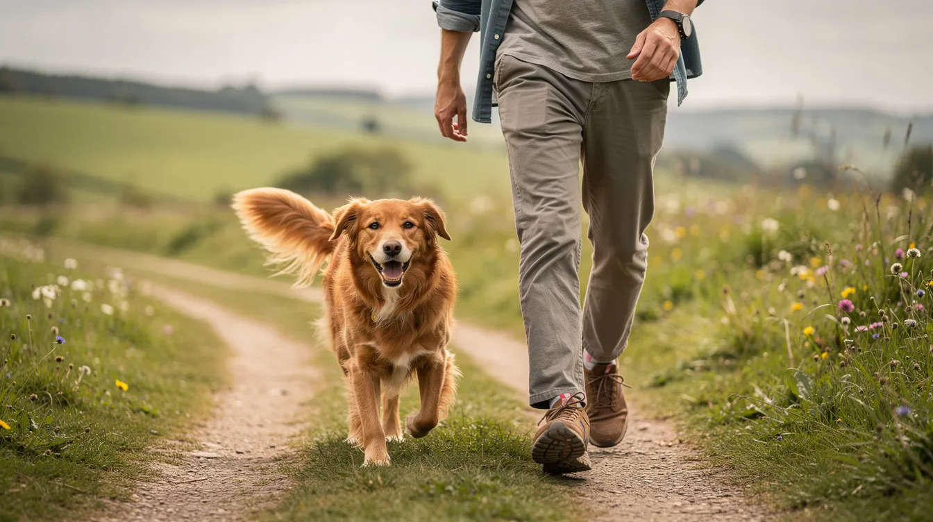
Aim for regular, moderate walks on even, non-slip surfaces. Multiple shorter outings (twenty to thirty minutes, two to three times daily) are generally better than one long walk. In wet or icy weather, be cautious - slippery surfaces increase the risk of jarring the affected elbow joints.
Appropriate activities:
- Steady lead walks on soft ground or grass
- Gentle off-lead mooching for suitable dogs on safe terrain
- Controlled swimming or hydrotherapy sessions
- Scentwork, puzzle feeders and training games for mental stimulation without physical impact
Activities to limit or avoid:
- Repetitive ball throwing and high-speed chasing
- Jumping in and out of cars or onto furniture
- Agility involving sharp turns or high jumps
- Rough play with significantly larger or boisterous dogs
Maintaining muscle mass
Consistent, moderate exercise combined with physiotherapy-guided strengthening is essential. Strong muscles around the shoulder and core help support the elbow and reduce loading on damaged structures.
Helpful adaptations at home:
- Ramps at doors, steps and into the car
- Non-slip rugs and runners over hard flooring
- Supportive orthopaedic beds in warm, draught-free areas
- Clear walkways free of obstacles
- Harnesses rather than collars to reduce strain on the neck and shoulders
Monitoring quality of life
Consider keeping a simple diary or using a scoring system to track your dog's comfort, mobility, appetite and enjoyment over time. Share this with your vet at regular reviews - it helps both of you make informed decisions about adjustments to the treatment plan.

Prevention, screening and responsible breeding
Elbow dysplasia cannot always be prevented, but risk can be meaningfully reduced through responsible breeding practices, appropriate growth management and early screening.
UK elbow scoring
The BVA/KC Elbow Dysplasia Scheme grades dogs from 0 to 3, with higher grades indicating more severe radiographic changes. Radiographs are taken after skeletal maturity (usually from twelve months of age). Breeders can use results from both parents to select lower-risk mating pairs, and long-term UK data shows gradual improvement in elbow grades over time, though progress has been slower than for hip dysplasia.
What to ask your breeder:
- Elbow scores for both parents
- Health testing results for the breed
- How puppies are raised - nutrition, exercise, socialisation
Preventing excessive weight gain during growth
- Choose a large-breed-specific complete diet
- Follow feeding guidelines carefully and avoid overfeeding
- Conduct regular weight checks - your vet can help assess body condition score
- Limit high-calorie treats and human food
Exercise advice for puppies at risk
Multiple short, controlled outings on safe surfaces are better than one long walk. Avoid repetitive "jump down" injuries from sofas, car boots or high steps. Limit stairs in the first six months of life.
Even with excellent breeding and careful management, some dogs will still develop elbow dysplasia because of the complex genetic basis of the condition. Early recognition and prompt veterinary care remain the most effective safety net.
How elbow dysplasia leads to osteoarthritis
Understanding why osteoarthritis is so closely linked to elbow dysplasia helps explain why lifelong management matters.
When the elbow joint is affected by any form of dysplasia - FCP, UAP, OCD or incongruity - the normal, even distribution of forces is disrupted. Instead of load spreading smoothly across the entire joint surface, abnormal pressure concentrates on small areas, particularly the medial compartment.
This concentrated loading damages the articular cartilage. Once the smooth cartilage surface is roughened, cracked or worn away, inflammation develops in the joint lining (synovitis). The body responds by producing new bone around the joint margins (osteophytes) and thickening the joint capsule. This progressive cycle of damage and attempted repair is osteoarthritis.
All dogs with elbow dysplasia develop elbow osteoarthritis to some degree. This process begins very early - sometimes before clinical signs are obvious - and continues throughout the dog's lifetime. Osteoarthritis progression is inevitable despite treatment, but the rate and severity can be significantly influenced by weight control, appropriate exercise, timely surgical intervention and long-term supportive care.
Think of it like a door hinge that is slightly misaligned. It still works, but it wears out much faster than a well-fitted hinge. The goal of management is to slow that wear and keep the "door" opening as smoothly as possible for as long as possible.
For a deeper understanding of osteoarthritis in dogs, visit our guides on Arthritis in Dogs and the Complete Guide to Joint Health in Dogs.
Graphic placeholder: How elbow dysplasia progresses to osteoarthritis - flow diagram from developmental lesion to chronic OA.
Myths and facts about elbow dysplasia
Myth: Only giant breed dogs develop elbow dysplasia. Fact: While giant breeds are over-represented, medium-sized breeds such as English Springer Spaniels and even some smaller dogs and crossbreeds can be affected.
Myth: Puppies with elbow dysplasia will grow out of it. Fact: Elbow dysplasia is a developmental structural problem. Dogs do not grow out of it. Without management, it progresses to worsening osteoarthritis.
Myth: If my dog can still run, the elbows cannot be that bad. Fact: Dogs are excellent at masking pain. Willingness to run does not mean the elbows are healthy. Many dogs with significant cartilage loss still attempt normal activity.
Myth: Surgery cures arthritis. Fact: Surgical intervention addresses the primary lesion (fragment, loose process, incongruity) but cannot reverse existing arthritis. It aims to slow progression and improve comfort.
Myth: Total rest is always the best treatment for front leg lameness. Fact: Prolonged rest leads to muscle loss and joint stiffness. Controlled, regular exercise is usually far more beneficial than complete rest.
Myth: Joint supplements can repair damaged cartilage completely. Fact: Supplements can support joint health and comfort but cannot regenerate lost cartilage or correct developmental abnormalities.
Myth: Front leg limping is always just a sprain. Fact: Persistent or recurring forelimb lameness in a young, large-breed dog warrants investigation for elbow dysplasia, not just rest and assumption of a soft tissue injury.
Myth: If X-rays are normal, it cannot be elbow dysplasia. Fact: Radiographs can miss subtle coronoid lesions and early incongruity. CT or arthroscopy may be needed for a definitive diagnosis.
Myth: If only one elbow looks sore, the other must be fine. Fact: Elbow dysplasia is bilateral in the majority of cases. Both elbows should be assessed even when only one seems clinically worse.
Myth: Painkillers alone are enough long-term management. Fact: Effective management combines weight control, exercise modification, physiotherapy, environmental adaptations and, where appropriate, supplements and surgical treatment. No single approach is sufficient on its own.
Graphic placeholder: Myth vs Fact infographic.
Frequently asked questions about elbow dysplasia
Can elbow dysplasia be cured? The underlying structural abnormality cannot be reversed. However, with appropriate treatment, most dogs achieve significantly improved comfort and function. The goal is management, not cure.
Does my dog need surgery? Not all dogs with elbow dysplasia require surgery. The decision depends on the type and severity of the lesion, your dog's age, degree of cartilage damage and response to conservative management. Your vet or orthopaedic specialist will advise on the best approach.
How much does elbow surgery cost in the UK? Costs vary widely. Arthroscopic fragment removal may range from £1,500 to £3,500 per elbow at referral centres. Osteotomy procedures and elbow replacement surgery are considerably more expensive. Pet insurance may cover some or all costs depending on your policy.
Will my dog get worse without surgery? The disease process tends to be progressive. Without appropriate management - whether surgical or conservative - cartilage damage and osteoarthritis generally worsen over time.
Can my dog still go for walks? Yes. Regular, controlled walks are an important part of management. Aim for shorter, frequent outings on even surfaces rather than long, strenuous hikes.
Is hydrotherapy safe for dogs with elbow dysplasia? Hydrotherapy is generally very beneficial, providing exercise with reduced joint impact. Always seek a vet referral to a reputable hydrotherapy centre experienced with orthopaedic patients.
How long will my dog need pain relief? This varies. Some dogs need ongoing NSAID therapy; others manage well with periodic courses during flare-ups, combined with supplements and lifestyle management.
Can I insure a dog with elbow dysplasia? Most insurers will not cover pre-existing conditions. If your dog is insured before diagnosis, treatment may be covered depending on your policy terms. Always check with your insurer.
Is elbow dysplasia hereditary? Yes. Genetic predisposition plays a major role in development. Breeding from dogs with low elbow scores reduces - but does not eliminate - risk in offspring.
Should I breed from a dog with elbow dysplasia? Most breed health experts and the BVA/KC scheme advise against breeding from dogs with high elbow grades. Responsible breeding is one of the most effective long-term strategies for reducing prevalence.
Will problems return after surgery? Surgery addresses the primary lesion, but secondary osteoarthritis will continue to develop over time. Ongoing management - weight control, exercise, supplements, regular veterinary reviews - remains essential.
Can small dogs get elbow dysplasia? Although elbow dysplasia predominantly affects medium, large and giant breeds, it can occasionally occur in smaller dogs. Persistent front leg lameness in any breed should be investigated.
How do I know if my dog is in pain? Dogs rarely cry out with chronic pain. Look for subtle signs: reluctance to exercise, stiffness after rest, licking at joints, changes in behaviour, reduced appetite or withdrawal from interaction.
What is the prognosis for a dog with elbow dysplasia? Prognosis depends on the type and severity of the lesion, age at diagnosis and quality of ongoing care. With appropriate treatment, many dogs enjoy an excellent quality of life for many years.
Can diet or supplements stop elbow dysplasia? No supplement or diet can prevent or cure elbow dysplasia. However, a balanced diet during growth reduces risk, and joint supplements can support comfort as part of long-term osteoarthritis management.
What is the difference between elbow dysplasia and simple arthritis? Elbow dysplasia refers to the developmental abnormality that causes abnormal joint loading. Arthritis (osteoarthritis) is the progressive joint degeneration that develops as a consequence. Treatment addresses both the primary problem and its secondary effects.
Do dogs respond well to treatment overall? Yes - most dogs respond positively to a combination of treatment approaches. Early diagnosis and proactive management give the best results.
Elbow dysplasia vs other causes of front leg lameness
While elbow dysplasia is a very common cause of forelimb lameness in young, medium and large breeds, other conditions can produce similar signs.
Other common causes of front leg limping:
- Shoulder conditions - osteochondrosis of the shoulder, biceps tendinopathy, or instability
- Carpal (wrist) injuries - sprains, fractures or hyperextension
- Soft tissue strains - muscle or ligament injuries in the leg or chest
- Trauma or fractures - from falls, road traffic incidents or play injuries
- Nerve problems - brachial plexus injuries
- Neck or spinal disease - disc problems causing referred forelimb pain
Accurate diagnosis matters because treatment plans differ significantly between these conditions. You are not expected to distinguish them at home - this is the role of your veterinary team. A thorough clinical examination and appropriate diagnostic imaging will determine the cause and guide the right approach.
Graphic placeholder: Decision tree - "My dog is limping on a front leg - what should I do?" guiding owners towards timely veterinary assessment.
Working with your vet and the Alder & Hound Knowledge Centre
Managing elbow dysplasia is a long-term partnership between you, your primary vet, any referral orthopaedic specialist, and allied professionals such as physiotherapists, hydrotherapists and nutritionists.
Practical tips for getting the most from veterinary appointments:
- Keep a simple lameness diary noting good and bad days
- Record short videos of your dog walking and trotting on different surfaces and days - these are invaluable for vets assessing progress
- Write down your questions before each appointment
- Note any response (positive or negative) to rest, medication or exercise changes
The Alder & Hound Canine Mobility Knowledge Centre is designed to support you between appointments. You will find evidence-informed guides covering related topics including hip dysplasia, cruciate ligament disease, arthritis in dogs, joint stiffness, and ingredient-specific guides for the supplements commonly used in joint health.
Alder & Hound provides educational content first and foremost, alongside premium joint-health supplements like ForeverRun™ that can be discussed with your vet as part of a broader, evidence-informed management plan.
Sign up for email updates to receive new educational articles, practical resources and occasional promotions - all focused on helping your dog stay comfortable, active and enjoying life.
Key takeaways: helping your dog stay comfortable and active
- Elbow dysplasia is a developmental condition - an umbrella term for four distinct joint abnormalities - and not a single disease
- It is the most common cause of front leg lameness in young, medium and large breed dogs
- Early diagnosis gives the best chance of slowing osteoarthritis progression and maintaining comfort
- Weight control is the single most impactful thing you can do at home
- Controlled exercise, physiotherapy and environmental adaptations protect the joint day to day
- Surgery can significantly improve comfort but does not cure arthritis - realistic expectations matter
- Joint supplements support long-term joint health but do not replace veterinary treatment
- Lifelong, multimodal management allows many dogs to live active, happy lives for many years
You know your dog best. Noticing subtle changes early - a slight stiffness, a shortened stride, a reluctance to play - and seeking timely veterinary advice is one of the most powerful things you can do. With the right care, support and information, dogs affected by elbow dysplasia can enjoy an excellent quality of life.
Continue learning through the Alder & Hound Canine Mobility Knowledge Centre - your trusted resource for evidence-informed canine joint health.